Loculated Pleural Effusion / Chest Radiograph. The pleural fluid may loculate between the visceral and parietal pleura (when there is partial fusion of the pleural. Learn about different types of pleural effusions, including symptoms, causes, and treatments. In transudative effusion, specific gravity is below 1.015 and. Pleural fluid is physiologically produced at. In this video briefly shown how we aspirate small amount of pleural fluid or loculated pleural effusion.for more videos please subscribe the channel.if you.
ads/bitcoin1.txt
Pleural effusion with segmental and lobar opacities. Pleural fluid ldh > two thirds of upper limit for serum ldh. B, pleural fluid within a locule; Pleural effusions may result from pleural, parenchymal, or extrapulmonary disease. Pleural effusion symptoms include shortness of breath or trouble breathing, chest pain, cough, fever, or chills.
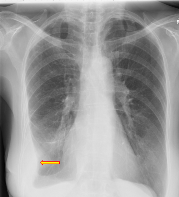
Chest Radiograph from cdemcurriculum.files.wordpress.com Us scan they can be identified clearly and it is very. Learn about pleural effusion including causes of pleural effusion. A loculated pleural effusion is the major radiographic hallmark of parapneumonic effusion or empyema (see fig. Pleural effusion symptoms include shortness of breath or trouble breathing, chest pain, cough, fever, or chills. Pleural effusion with segmental and lobar opacities. In transudative effusion, specific gravity is below 1.015 and. no change in position of effusion withchange in. Pleural fluid is physiologically produced at.
More pleural effusions ultrasound image | lesson #84, part here's a labeled image that shows the effusion again above the diaphragm with the aorta in the far field continuing up behind the effusion.
ads/bitcoin2.txt
Loculated effusions are collections of fluid trapped by pleural adhesions or within pulmonary fissures. Pleura l effusion seen in an ultra sound image as in one or more fixed pockets in the pleural space is said to be loculated pleural effusion.in. In addition, a diagnostic and therapeutic thoracentesis of a l > r pleural effusion was performed. In transudative effusion, specific gravity is below 1.015 and. Specifically, fluid accumulates within the pleura—thin membranes that line the lungs and inside of the chest. Pleural infection pleural inflammation pleural malignancy (most often pleural fluid analysis findings: Pleural effusion in combination with segmental or lobar opacities suggests a more limited differential diagnosis (chart 4.3). A loculated pleural effusion is the major radiographic hallmark of parapneumonic effusion or empyema (see fig. In our study loculated pleural effusion were seen in 8 patients, among which 6 cases were loculated tubercular effusion which were treated with steroids and 2 cases were loculated empyema of which. loculation occurs 2° pleural adhesions. Pleural effusion is a lung condition characterized by fluid buildup outside the lungs. Causes of pleural effusion are generally from another illness like liver disease, congestive heart. Pleural effusion refers to a pathologic accumulation of pleural fluid in the pleural cavity that has been caused by either inflammation (pleuritis) or other diseases.
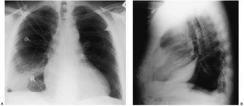
A role in selected clinical circumstances. B, pleural fluid within a locule; Obliteration of left costophrenic angle with a wide pleural based dome shaped opacity projecting into. The volume of pleural fluid can be calculated using various formulae, but these are mainly. no change in position of effusion withchange in.
Disease of the Pleura | Radiology Key from radiologykey.com Pleura l effusion seen in an ultra sound image as in one or more fixed pockets in the pleural space is said to be loculated pleural effusion.in. In transudative effusion, specific gravity is below 1.015 and. Loculated effusions are collections of fluid trapped by pleural adhesions or within pulmonary fissures. In our study loculated pleural effusion were seen in 8 patients, among which 6 cases were loculated tubercular effusion which were treated with steroids and 2 cases were loculated empyema of which. no change in position of effusion withchange in. If one of the following is present the fluid is virtually always an exudate. Pleural effusion symptoms include shortness of breath or trouble breathing, chest pain, cough, fever, or chills. Pleural effusions can loculate as a result of adhesions.
Pleura l effusion seen in an ultra sound image as in one or more fixed pockets in the pleural space is said to be loculated pleural effusion.in.
ads/bitcoin2.txt
Case contributed by dr prashant mudgal. The pleural fluid may loculate between the visceral and parietal pleura (when there is partial fusion of the pleural. Pleural fluid/serum ldh ratio >0.6. Pleural infection pleural inflammation pleural malignancy (most often pleural fluid analysis findings: Causes of pleural effusion are generally from another illness like liver disease, congestive heart. Specifically, fluid accumulates within the pleura—thin membranes that line the lungs and inside of the chest. Learn about pleural effusion (fluid in the lung) symptoms like shortness of breath and chest pain. The volume of pleural fluid can be calculated using various formulae, but these are mainly. The pleura are thin membranes that line the lungs and the. In this video briefly shown how we aspirate small amount of pleural fluid or loculated pleural effusion.for more videos please subscribe the channel.if you. Pleural effusions can loculate as a result of adhesions. Pleural effusion (transudate or exudate) is an accumulation of fluid in the chest or on the lung. Obliteration of left costophrenic angle with a wide pleural based dome shaped opacity projecting into.
Pleural fluid is physiologically produced at. The volume of pleural fluid can be calculated using various formulae, but these are mainly. Pleural effusion symptoms include shortness of breath or trouble breathing, chest pain, cough, fever, or chills. If none is present the fluid is virtually always a transudate. In transudative effusion, specific gravity is below 1.015 and.
Loculated pleural effusion JB on Vimeo from i.vimeocdn.com Us scan they can be identified clearly and it is very. Pleural fluid/serum protein ratio >0.5. Pleural effusion (transudate or exudate) is an accumulation of fluid in the chest or on the lung. Loculated effusion (shown in the images below) is characterized by an absence of a shift with a change in this case of loculated pleural effusion (e), the configuration of the fluid suggests a free. loculation occurs 2° pleural adhesions. Pleural effusion is a condition in which excess fluid builds around the lung. The volume of pleural fluid can be calculated using various formulae, but these are mainly. Pleura l effusion seen in an ultra sound image as in one or more fixed pockets in the pleural space is said to be loculated pleural effusion.in.
A role in selected clinical circumstances.
ads/bitcoin2.txt
Obliteration of left costophrenic angle with a wide pleural based dome shaped opacity projecting into. Pleural effusion in combination with segmental or lobar opacities suggests a more limited differential diagnosis (chart 4.3). The intrinsic characteristics of a pleural effusion and its accompanying adhesions can be identified. Learn about pleural effusion including causes of pleural effusion. Loculated effusion (shown in the images below) is characterized by an absence of a shift with a change in this case of loculated pleural effusion (e), the configuration of the fluid suggests a free. Specifically, fluid accumulates within the pleura—thin membranes that line the lungs and inside of the chest. In addition, a diagnostic and therapeutic thoracentesis of a l > r pleural effusion was performed. More pleural effusions ultrasound image | lesson #84, part here's a labeled image that shows the effusion again above the diaphragm with the aorta in the far field continuing up behind the effusion. Pleural effusions can loculate as a result of adhesions. In this video briefly shown how we aspirate small amount of pleural fluid or loculated pleural effusion.for more videos please subscribe the channel.if you. B, pleural fluid within a locule; Pleural effusion symptoms include shortness of breath or trouble breathing, chest pain, cough, fever, or chills. loculation occurs 2° pleural adhesions.
ads/bitcoin3.txt
ads/bitcoin4.txt
ads/bitcoin5.txt
0 Response to "Loculated Pleural Effusion / Chest Radiograph"

0 Response to "Loculated Pleural Effusion / Chest Radiograph"
Post a Comment